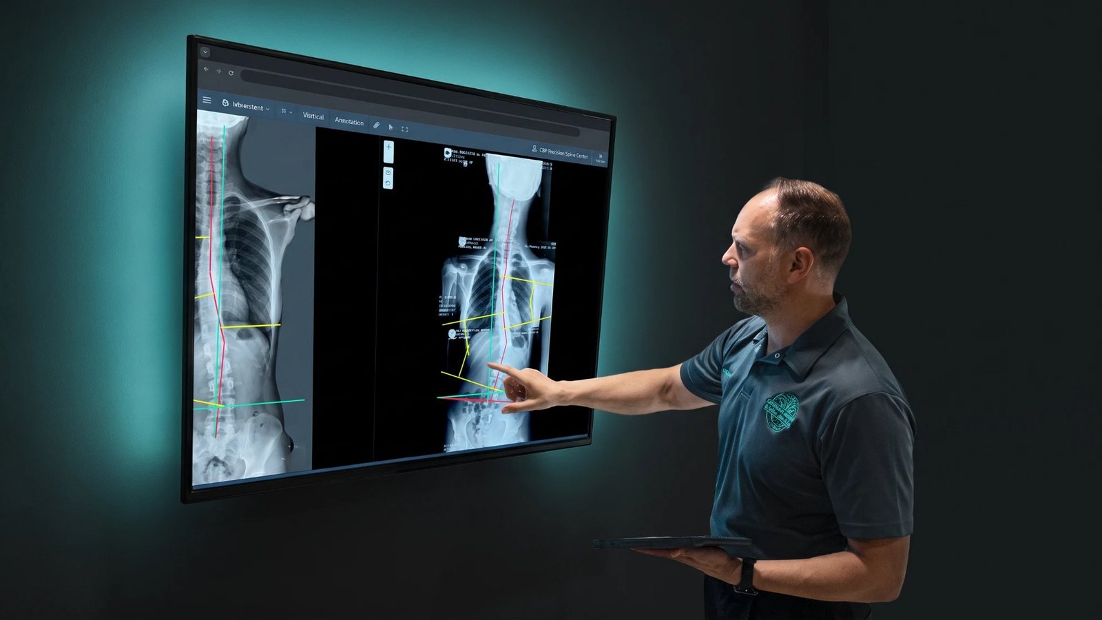
Most scoliosis stories start the same way. A parent notices one shoulder sitting higher than the other, or a swimming coach mentions that a child's back looks uneven when they bend forward. An adult is told at a routine check that their spine "has a bit of a curve". And then, far too often, comes the next sentence: "Let's just keep an eye on it."
Keep an eye on what, exactly? Nobody can see a spine through skin. You can see a shoulder, a hip, a rib hump. Those are clues. The spine itself (how far it bends, how much it twists, where the head sits over the pelvis, whether the neck and lower back still have their normal curves) only shows up on a standing X-ray.
At CBP Precision Spine Center we practise Chiropractic BioPhysics® (CBP), a structural approach to spine care. Structural care has one simple rule: measure the spine before you try to change it, then measure it again to prove whether it changed. No film, no measurement. No measurement, no honest plan. This guide explains when you need scoliosis X-rays, why they must be taken standing (from the front and the side), how the radiation question really stacks up, and what the numbers on your report mean.
Does every curve need an X-ray?
If there is a real sign of a curve, yes. Uneven shoulders, a hip that sits higher, a waist that looks different on each side, or a rib hump when bending forward are not "just posture" until somebody proves it. They are signs, and the only way to prove what is causing them is to look at the spine itself.
The examination still comes first, every time. We look at how you stand and move, do the forward-bend test with a scoliometer, measure your posture, check leg length and take a proper history: when the change was first noticed, whether it is getting worse, and whether anyone else in the family has scoliosis. We have written before about why we examine first and image second. But "examine first" does not mean "never image". The exam tells us where to look. The X-ray tells us what is actually there.
Here is the problem with stopping at the exam. A forward-bend test and a posture photo cannot give you a Cobb angle. They cannot show how much each vertebra is rotated, whether the curve is structural or postural, or what the neck and lower back are doing from the side. Two people can look almost identical from the outside and have very different spines. "It looks mild" is an opinion. "It measures 18°" is a fact.
What about "just posture"? A side-to-side curve under 10° on a standing film is not called scoliosis; it is most likely postural, and a postural shift can usually be improved back towards normal. But notice the catch: you only know it is under 10° because someone measured it on a film. And posture problems are spinal problems too. Forward head posture, a lost neck curve, a shifted ribcage or a tilted pelvis are changes in spinal alignment, and in CBP they are measured on X-ray exactly the way a scoliosis is.
So we arrange standing X-rays when:
- the examination finds a sign of a curve or an alignment problem;
- we are considering corrective (structural) care, because we will not plan to correct a spine we have not seen;
- we need a baseline to measure your progress against;
- something in the history or exam needs ruling out before we adjust or apply traction.
We do not arrange them when the examination is clear, when you already have recent standing films we can use, or during pregnancy. We do not X-ray children routinely either, but a child with a positive screening test is not routine. For a growing child, finding the curve early is the whole game.
Why a scoliosis X-ray is taken standing
A proper scoliosis study is a full-spine X-ray, from the base of the skull to the pelvis, taken while you stand. And it is two views, not one: one from the front and one from the side. Standing matters for a reason you can feel yourself. Lying down takes your body weight off the spine, so a curve usually measures smaller lying down than standing. You do not live lying down. The standing film shows your spine under your own weight, the way you sit, walk and work every day.
The side view matters just as much, and it is the one most often skipped. Scoliosis is not a flat, side-to-side bend; it is a three-dimensional twist. From the side we can see whether the mid-back has lost its normal rounding (common in adolescent scoliosis), whether the neck and lower back have kept their curves, and whether the head and trunk sit balanced over the pelvis. A front view alone gives you half the picture and a confident-sounding report.
Standing films are also repeatable. A follow-up film taken in the same position, with the same set-up, can be compared fairly, and a real change then means something. Comparing a standing film with a lying-down one means very little, which is why we will still ask for standing films even if you bring older ones taken lying down.
What happens at the imaging centre
The appointment itself is short. We refer you to an imaging centre for full-spine standing X-rays, front and side, and analyse the films here. The radiographer positions you against the detector, asks you to stand naturally with your arms out of the way, and takes the views, each lasting a fraction of a second. You will be asked to remove jewellery, belts and anything metal in the area, because metal blocks the image.
It helps to bring:
- the referral we gave you, which names the exact views we need;
- for a child, a familiar adult who can stand nearby.
Helping a child through it
Children do best with a calm, truthful description: "They will take a picture of your back. You stand still for a few seconds, and it doesn't hurt." Do not promise that nothing will feel strange; the room can be cool and the plate is hard. Practise standing still for ten seconds at home the night before, and tell the imaging team when booking if your child has sensory, communication or mobility needs, so they can allow extra time.
The radiation question, answered honestly
Yes, X-rays use ionising radiation. No, a standing spine film is not the danger it is often made out to be. The dose from a modern digital spine study is low, roughly in the range of the natural background radiation all of us absorb over a few months just by living on Earth. It is a funny thing: a single standing spine film tends to cause more worry than a CT scan, which delivers many times the dose and is ordered far more casually. Researchers in our own field have written about this "radiophobia" and why it should not stop spine and posture patients from being properly imaged (Oakley & Harrison, Dose-Response, 2018).
The risk people rarely talk about is the risk of not taking the film: a curve that is missed, a structural problem treated as "just posture", a plan built on guesswork, or a growing child's curve that progresses while everyone keeps an eye on it. Those risks are real, and for a growing spine they cannot be undone later.
That said, every film still has to earn its place. We keep exposure down in practical ways:
- Image when the result changes the plan. No film "just to see", and no plan without one.
- Do not repeat what already exists. A recent standing film from another provider is often usable. Bring the images, not only the report.
- Re-image at set points, timed by the clinical question (for a growing child, how fast the curve is changing and how much growth is left), not by habit.
- Take the views that answer the question, and nothing extra.
Reading the report: the Cobb angle and the rest
A radiologist's report and our analysis are not the same thing. The report screens for disease, fractures and anything unusual, and it often sums the curve up in a single line. Our analysis measures your alignment segment by segment, from the front and the side, and compares it with normal values. Here is what we look at:
| Item | What it describes | Why it matters |
|---|---|---|
| Curve location | Which part of the spine curves (mid-back, lower back, or both) | Different curve patterns behave differently and need different care |
| Curve direction | Whether the curve bends to the right or to the left | Sets the direction of every corrective exercise, traction set-up and brace |
| Cobb angle | The size of the curve in degrees | The standard number used to grade severity and track change |
| Vertebral rotation | How much the vertebrae twist within the curve | Rotation drives the rib hump and how the curve may progress |
| Side-view curves | The curves of the neck, mid-back and lower back seen from the side | Loss of these curves changes how the spine carries load, and is part of the correction |
| Spinal balance | Whether the head and trunk sit over the pelvis, front and side | Affects symptoms and function as much as curve size |
| Pelvis and leg length | Whether the pelvis is level | An unlevel base can drive or worsen a curve |
| Growth markers | How much skeletal growth is left (children and teenagers) | Growth remaining is the strongest guide to how fast a curve may progress |
The Cobb angle is the angle between the most tilted vertebra at the top of the curve and the most tilted vertebra at the bottom. By international convention a curve of 10° or more on a standing film is scoliosis. The SOSORT guidelines grade it like this:
| Cobb angle | SOSORT grade |
|---|---|
| Under 10° | Not scoliosis (most likely postural) |
| 10° to 20° | Low (mild) |
| 21° to 35° | Moderate |
| 36° to 40° | Moderate to severe |
| 41° to 50° | Severe |
| 51° to 55° | Severe to very severe |
| 56° and over | Very severe |
Our non-surgical scoliosis guide explains what each grade usually means in practice. One honest caveat: two readings of the same film can differ by a few degrees, so a change of 2° or 3° between films is within the margin of error. That is exactly why we measure with the same computer-assisted method every time, and why we read the trend rather than any single number.
What the film cannot tell you
An X-ray shows structure. It does not show pain, stiffness, how far you can bend, how long you can stand, or how you feel about your back. Scoliosis can hurt, but it does not hurt everyone equally: some people with a small curve are very sore and some with a larger one feel fine. That is why the film never replaces the examination. The exam tells us how your spine functions; the film tells us how it is built and aligned. You need both, and anyone offering you a plan with only one of them is guessing at the other half.
What we do with your films
Bring your X-rays to your appointment on disc or as digital files, with the report if you have one. We analyse the standing films here, measure the Cobb angle, rotation, side-view curves and balance, and put those numbers next to your posture measurements and examination findings. Then we sit down with you, show you your own spine on screen, and explain what we see. Only then do we talk about a plan.
For scoliosis, a corrective plan at our clinic is built around the two things that do the heavy lifting: scoliosis-specific Mirror Image® exercise and corrective traction. Chiropractic adjustments support mobility and comfort, and where the curve, age and growth call for it, we fit a custom ScoliBrace®. For a growing child, we start scoliosis-specific exercise as early as possible rather than simply watching, in line with SOSORT and ScoliCare recommendations.
And we do not ask you to take our word for it. We re-examine and re-image at set points, taken the same way as the first films, so any claim of progress rests on a before-and-after comparison you can see for yourself. You can read more about the method on our Chiropractic BioPhysics® page and the full picture on our scoliosis assessment page.
Five questions to ask before any scoliosis X-ray
- Will it be taken standing, and will it show the whole spine?
- Will I get both a front view and a side view?
- Who will measure it, and will the same method be used next time?
- Will I get the actual images, not just the written report?
- If I am told I do not need one, how will the curve be measured instead?
A clinician who answers those five clearly is one you can work with. For more on judging a provider, see how to choose spine care in Muscat, and if the patient is a child, our guide to scoliosis screening in children.
Frequently asked questions
Does a scoliosis X-ray hurt, and how long does it take?
It does not hurt. You stand still for a few seconds per view, and the whole visit, including positioning and paperwork, usually takes well under half an hour. Children sometimes find the cool room and the hard plate unfamiliar, which is why a calm explanation beforehand helps.
Can scoliosis be checked without an X-ray?
It can be screened without one. The forward-bend test, shoulder and hip levels and posture measurements tell us whether a curve is likely. But it cannot be confirmed, measured or tracked without a standing film. If there is a sign on examination, the film is the next step, not an optional extra.
Isn't it safer to just wait and watch?
Watching a curve is not the same as treating it. For a growing child, months of waiting can mean months of progression during the very window when care works best. We would rather measure it, know exactly where we stand, and act early.
My earlier X-ray was taken lying down. Is it still useful?
Bring it. It still shows the location and shape of the curve. But curves usually measure smaller lying down, so for a baseline to monitor or correct against, we will need standing films.
How often does a scoliosis X-ray need repeating?
It depends on age, growth and the plan. A growing child in active care is usually re-imaged every 6 months; an adult with a stable curve far less often. We also re-image at the end of a corrective phase to measure the result, and we always take the films the same way so the comparison is fair.
Do I get the X-ray before or after my first appointment?
Usually after. We examine you first, decide which views are needed, then refer you for standing X-rays and go through them with you at your next visit. If you already have recent standing films, bring them to the first appointment and you may not need new ones.
What counts as a normal Cobb angle?
A side-to-side curve under 10° on a standing film is not called scoliosis. It is most likely postural, and it can usually be improved closer to normal. From 10° upwards it is graded using the SOSORT scale above. The number alone does not decide treatment; age, growth remaining, balance and symptoms matter as much.
If you or your child has been told to "keep an eye on" a curve, or you already have films you would like read properly, book an assessment at CBP Precision Spine Center, 336 18th November Street, Azaiba. Call 7277 7796 or book online. Live outside Muscat? Send us your standing X-rays and posture photos and we will review them for you.
تبدأ معظم قصص الجنف بالطريقة نفسها. تلاحظ الأم أنّ كتف طفلها أعلى من الأخرى، أو يذكر مدرّب السباحة أنّ ظهر الطفل يبدو غير متناظر حين ينحني إلى الأمام. أو يُقال لشخص بالغ في فحص روتيني إنّ في عموده الفقري «اعوجاجاً بسيطاً». ثم تأتي، في أحيان كثيرة جداً، الجملة التالية: «لنراقبه فقط».
نراقب ماذا بالضبط؟ لا أحد يرى العمود الفقري من خلال الجلد. ترى الكتف والورك وبروز الأضلاع، وهذه كلّها مؤشّرات. أمّا العمود الفقري نفسه (إلى أيّ مدى ينحني، وكم يلتفّ، وأين يقع الرأس فوق الحوض، وهل ما زالت الرقبة وأسفل الظهر يحتفظان بانحناءاتهما الطبيعية) فلا يظهر إلا في صورة أشعة تُؤخذ واقفاً.
في مركز الدقة للعمود الفقري نمارس الكيروبراكتيك البيوفيزيائي Chiropractic BioPhysics® (CBP)، وهو منهج بنيوي في رعاية العمود الفقري. وللرعاية البنيوية قاعدة بسيطة واحدة: قِس العمود الفقري قبل أن تحاول تغييره، ثم قِسه مرة أخرى لتُثبت هل تغيّر. لا صورة، لا قياس. ولا قياس، لا خطة صادقة. يشرح هذا الدليل متى تحتاج إلى صور أشعة الجنف، ولماذا يجب أن تُؤخذ واقفاً (من الأمام ومن الجانب)، وحقيقة سؤال الإشعاع، وماذا تعني الأرقام في تقريرك.
هل يحتاج كلّ اعوجاج إلى صورة أشعة؟
إذا كانت هناك علامة حقيقية على انحناء، فنعم. الكتفان غير المتساويين، أو الورك الأعلى من الآخر، أو الخصر الذي يبدو مختلفاً بين الجانبين، أو بروز الأضلاع عند الانحناء إلى الأمام، ليست «مجرّد وضعية» حتى يُثبت أحدٌ ذلك. إنها علامات، والطريقة الوحيدة لإثبات سببها هي النظر إلى العمود الفقري نفسه.
ومع ذلك يأتي الفحص أولاً في كلّ مرة. ننظر في طريقة وقوفك وحركتك، ونُجري اختبار الانحناء إلى الأمام بمقياس الجنف، ونقيس قوامك، ونفحص طول الساقين، ونأخذ تاريخاً مرضياً وافياً: متى لوحظ التغيّر أول مرة، وهل يزداد، وهل في العائلة من لديه جنف. وقد كتبنا من قبل لماذا نفحص أولاً ونصوّر ثانياً. لكنّ «الفحص أولاً» لا يعني «لا تصوير أبداً». الفحص يخبرنا أين ننظر، والصورة تخبرنا بما هو موجود فعلاً.
وهذه مشكلة التوقّف عند الفحص: اختبار الانحناء إلى الأمام وصورة القوام لا يعطيانك زاوية كوب. ولا يُظهران مقدار دوران كلّ فقرة، ولا هل الانحناء بنيوي أم وضعي، ولا ما يحدث في الرقبة وأسفل الظهر من الجانب. قد يبدو شخصان متطابقين تقريباً من الخارج ولديهما عمودان فقريان مختلفان جداً. «يبدو خفيفاً» رأي، و«قياسه 18°» حقيقة.
وماذا عن «مجرّد وضعية»؟ الانحناء الجانبي الذي يقلّ عن 10° في الصورة واقفاً لا يُسمّى جنفاً؛ فهو على الأرجح وضعي، ويمكن عادةً تحسين الانزياح الوضعي ليعود نحو الطبيعي. لكن انتبه إلى النقطة المهمّة: أنت تعرف أنه أقلّ من 10° لأنّ أحداً قاسه على صورة. ثم إنّ مشكلات القوام مشكلات في العمود الفقري أيضاً. تقدّم الرأس، وفقدان انحناء الرقبة، وانزياح القفص الصدري، وميلان الحوض، كلّها تغيّرات في استقامة العمود الفقري، وفي منهج CBP تُقاس على الأشعة تماماً كما يُقاس الجنف.
لذلك نرتّب صور أشعة واقفاً حين:
- يجد الفحص علامة على انحناء أو مشكلة في الاستقامة؛
- نفكّر في رعاية تصحيحية (بنيوية)، لأننا لن نخطّط لتصحيح عمود فقري لم نره؛
- نحتاج إلى خطّ أساس نقيس تقدّمك عليه؛
- يحتاج أمرٌ في التاريخ المرضي أو الفحص إلى استبعاد قبل التعديل أو الشدّ.
ولا نرتّبها حين يكون الفحص سليماً، أو حين تكون لديك صور حديثة واقفاً يمكننا استخدامها، أو أثناء الحمل. كما أننا لا نصوّر الأطفال بالأشعة روتينياً، لكنّ الطفل الذي جاءت نتيجة فحصه إيجابية ليس حالة روتينية. وعند الطفل في طور النمو، اكتشاف الانحناء مبكراً هو كلّ شيء.
لماذا تُؤخذ صورة أشعة الجنف واقفاً؟
دراسة الجنف الصحيحة صورة أشعة للعمود الفقري كاملاً، من قاعدة الجمجمة إلى الحوض، تُلتقط وأنت واقف. وهي لقطتان لا واحدة: واحدة من الأمام وأخرى من الجانب. والوقوف مهمّ لسبب تشعر به بنفسك: الاستلقاء يرفع وزن جسمك عن العمود الفقري، فيبدو الانحناء عادةً أصغر في القياس وأنت مستلقٍ منه وأنت واقف. وأنت لا تعيش مستلقياً. الصورة واقفاً تُظهر عمودك الفقري تحت وزنك أنت، كما تجلس وتمشي وتعمل كلّ يوم.
واللقطة الجانبية مهمّة بالقدر نفسه، وهي الأكثر تجاهلاً. فالجنف ليس انحناءً مسطّحاً من جانب إلى جانب؛ إنه التواء ثلاثي الأبعاد. ومن الجانب نرى هل فقد منتصف الظهر استدارته الطبيعية (وهذا شائع في جنف المراهقين)، وهل احتفظت الرقبة وأسفل الظهر بانحناءاتهما، وهل يقع الرأس والجذع في توازن فوق الحوض. اللقطة الأمامية وحدها تعطيك نصف الصورة وتقريراً يبدو واثقاً.
والصور واقفاً قابلة للتكرار أيضاً. فصورة المتابعة المأخوذة في الوضعية نفسها، وبالإعداد نفسه، يمكن مقارنتها مقارنة عادلة، وعندها يعني التغيّر الحقيقي شيئاً. أمّا مقارنة صورة واقفاً بأخرى مستلقياً فلا تكاد تعني شيئاً، ولهذا سنطلب صوراً واقفاً حتى لو أحضرت صوراً أقدم أُخذت مستلقياً.
ماذا يحدث في مركز التصوير؟
الموعد نفسه قصير. نحوّلك إلى مركز تصوير لصور أشعة للعمود الفقري كاملاً واقفاً، من الأمام ومن الجانب، ثم نحلّل الصور هنا. يضعك فنّي الأشعة أمام اللوح، ويطلب منك أن تقف وقفة طبيعية وذراعاك بعيدتان عن مجال الصورة، ويلتقط اللقطات، ويستغرق كلّ منها جزءاً من الثانية. وسيُطلب منك نزع المجوهرات والحزام وأيّ شيء معدني في منطقة التصوير، لأنّ المعدن يحجب الصورة.
يفيدك أن تُحضر:
- ورقة التحويل التي أعطيناك إياها، وفيها اللقطات التي نحتاجها بالضبط؛
- وللطفل، شخصاً بالغاً يعرفه يستطيع الوقوف قريباً منه.
كيف تساعد طفلك على اجتيازها
يتجاوب الأطفال أفضل ما يتجاوبون مع وصف هادئ وصادق: «سيأخذون صورة لظهرك. تقف ثابتاً بضع ثوانٍ، ولا يؤلم». لا تعِد طفلك بأنّ شيئاً لن يبدو غريباً؛ فقد تكون الغرفة باردة واللوح صلب. تدرّبا على الوقوف ثابتين عشر ثوانٍ في البيت في الليلة السابقة، وإذا كان لدى طفلك احتياجات حسّية أو تواصلية أو حركية فأخبر فريق التصوير عند الحجز ليتيح وقتاً إضافياً.
سؤال الإشعاع، بإجابة صادقة
نعم، تستخدم الأشعة السينية إشعاعاً مؤيّناً. ولا، صورة العمود الفقري واقفاً ليست الخطر الذي تُصوَّر به غالباً. فالجرعة من دراسة رقمية حديثة للعمود الفقري منخفضة، وهي تقريباً في حدود الإشعاع الطبيعي الذي نمتصّه جميعاً خلال بضعة أشهر لمجرّد أننا نعيش على الأرض. والمفارقة أنّ صورة واحدة للعمود الفقري واقفاً تثير قلقاً أكثر مما تثيره الأشعة المقطعية (CT)، مع أنّ جرعتها أكبر بأضعاف وتُطلب بسهولة أكبر بكثير. وقد كتب باحثون في مجالنا عن هذا «الرهاب من الإشعاع» ولماذا لا ينبغي أن يمنع مرضى العمود الفقري والقوام من التصوير الصحيح (Oakley & Harrison، مجلة Dose-Response، 2018).
أمّا الخطر الذي نادراً ما يُتحدَّث عنه فهو خطر عدم أخذ الصورة: انحناء يفوت، أو مشكلة بنيوية تُعامَل على أنها «مجرّد وضعية»، أو خطة مبنية على التخمين، أو انحناء طفل في طور النمو يتفاقم بينما يكتفي الجميع بمراقبته. هذه مخاطر حقيقية، وفي العمود الفقري النامي لا يمكن تداركها لاحقاً.
ومع ذلك، يجب أن تستحقّ كلّ صورة مكانها. نُبقي التعرّض منخفضاً بطرق عملية:
- التصوير حين تغيّر النتيجة الخطة. لا صورة «لمجرّد الاطمئنان»، ولا خطة من دون صورة.
- عدم تكرار ما هو موجود أصلاً. الصورة الحديثة واقفاً من جهة أخرى تصلح في أحيان كثيرة. أحضر الصور نفسها، لا التقرير وحده.
- إعادة التصوير في نقاط محدّدة، يحدّد توقيتها السؤال السريري (عند الطفل في طور النمو: ما سرعة تغيّر الانحناء وكم بقي من النمو)، لا العادة.
- أخذ اللقطات التي تجيب عن السؤال، ولا شيء زائداً.
قراءة التقرير: زاوية كوب وما سواها
تقرير طبيب الأشعة وتحليلنا ليسا الشيء نفسه. التقرير يبحث عن الأمراض والكسور وأيّ شيء غير عادي، وكثيراً ما يلخّص الانحناء في سطر واحد. أمّا تحليلنا فيقيس استقامة عمودك الفقري جزءاً جزءاً، من الأمام ومن الجانب، ويقارنها بالقيم الطبيعية. وهذا ما ننظر إليه:
| البند | ماذا يصف | لماذا يهمّ |
|---|---|---|
| موضع الانحناء | أيّ جزء من العمود الفقري ينحني (منتصف الظهر، أو أسفل الظهر، أو كلاهما) | أنماط الانحناء المختلفة تسلك سلوكاً مختلفاً وتحتاج رعاية مختلفة |
| اتجاه الانحناء | هل ينحني إلى اليمين أم إلى اليسار | يحدّد اتجاه كلّ تمرين تصحيحي وإعداد للشدّ ودعامة |
| زاوية كوب | حجم الانحناء بالدرجات | الرقم المعياري لتصنيف الشدّة ومتابعة التغيّر |
| دوران الفقرات | مقدار التفاف الفقرات داخل الانحناء | الدوران هو ما يصنع بروز الأضلاع ويؤثّر في احتمال تفاقم الانحناء |
| الانحناءات الجانبية | انحناءات الرقبة ومنتصف الظهر وأسفله كما تُرى من الجانب | فقدانها يغيّر طريقة حمل العمود الفقري للأثقال، وهو جزء من التصحيح |
| توازن العمود الفقري | هل يقع الرأس والجذع فوق الحوض، من الأمام ومن الجانب | يؤثّر في الأعراض والوظيفة بقدر حجم الانحناء |
| الحوض وطول الساقين | هل الحوض مستوٍ | القاعدة غير المستوية قد تسبّب الانحناء أو تزيده سوءاً |
| مؤشّرات النمو | كم بقي من نموّ الهيكل العظمي (عند الأطفال والمراهقين) | النموّ المتبقّي أقوى دليل على سرعة تفاقم الانحناء المحتملة |
زاوية كوب هي الزاوية بين أكثر الفقرات ميلاً في أعلى الانحناء وأكثرها ميلاً في أسفله. وبالاصطلاح الدولي، الانحناء الذي يبلغ 10° أو أكثر في الصورة واقفاً جنف. وتصنّفه إرشادات SOSORT هكذا:
| زاوية كوب | درجة SOSORT |
|---|---|
| أقلّ من 10° | ليس جنفاً (على الأرجح وضعي) |
| من 10° إلى 20° | منخفض (خفيف) |
| من 21° إلى 35° | متوسّط |
| من 36° إلى 40° | متوسّط إلى شديد |
| من 41° إلى 50° | شديد |
| من 51° إلى 55° | شديد إلى شديد جداً |
| 56° فأكثر | شديد جداً |
ويشرح دليلنا لعلاج الجنف دون جراحة ما تعنيه كلّ درجة عملياً في العادة. وتنبيه صادق واحد: قد تختلف قراءتان للصورة نفسها ببضع درجات، فالتغيّر بـ2° أو 3° بين صورتين يقع ضمن هامش الخطأ. ولهذا بالضبط نقيس بالطريقة نفسها المدعومة بالحاسوب في كلّ مرة، ونقرأ الاتجاه العام لا رقماً واحداً بعينه.
ما الذي لا تستطيع الصورة أن تخبرك به
تُظهر صورة الأشعة البنية. ولا تُظهر الألم، ولا التيبّس، ولا إلى أيّ مدى تستطيع الانحناء، ولا كم تستطيع الوقوف، ولا كيف تشعر تجاه ظهرك. قد يؤلم الجنف، لكنه لا يؤلم الجميع بالقدر نفسه: بعض من لديهم انحناء صغير يتألّمون كثيراً، وبعض من لديهم انحناء أكبر لا يشعرون بشيء. ولهذا لا تحلّ الصورة أبداً محلّ الفحص. الفحص يخبرنا كيف يعمل عمودك الفقري، والصورة تخبرنا كيف هو مبنيّ ومستقيم. تحتاج إلى الاثنين، ومن يعرض عليك خطة بأحدهما فقط فهو يخمّن النصف الآخر.
ماذا نفعل بصورك
أحضر صور الأشعة إلى موعدك، على قرص أو ملفّات رقمية، مع التقرير إن وُجد. نحلّل الصور المأخوذة واقفاً هنا، ونقيس زاوية كوب والدوران والانحناءات الجانبية والتوازن، ونضع هذه الأرقام إلى جانب قياسات قوامك ونتائج فحصك. ثم نجلس معك، ونريك عمودك الفقري على الشاشة، ونشرح ما نراه. وعندها فقط نتحدّث عن الخطة.
وفي الجنف، تُبنى الخطة التصحيحية في مركزنا حول الركيزتين اللتين تؤدّيان العمل الأكبر: تمارين Mirror Image® المخصّصة للجنف والشدّ التصحيحي. وتدعم تعديلات الكيروبراكتيك الحركة والراحة، وحين يستدعي الانحناءُ والعمرُ والنموّ ذلك، نركّب دعامة ScoliBrace® مصنوعة خصيصاً. وعند الطفل في طور النمو، نبدأ التمارين المخصّصة للجنف في أبكر وقت ممكن بدلاً من الاكتفاء بالمراقبة، بما يتّفق مع توصيات SOSORT وScoliCare.
ولا نطلب منك أن تصدّقنا على كلامنا. نعيد الفحص والتصوير في نقاط محدّدة، بالطريقة نفسها التي أُخذت بها الصور الأولى، حتى يستند أيّ حديث عن تقدّم إلى مقارنة قبل وبعد تراها بعينك. يمكنك قراءة المزيد عن المنهج في صفحة الكيروبراكتيك البيوفيزيائي، والصورة الكاملة في صفحة تقييم الجنف.
خمسة أسئلة تطرحها قبل أيّ صورة أشعة للجنف
- هل ستُؤخذ واقفاً، وهل ستُظهر العمود الفقري كاملاً؟
- هل سأحصل على لقطة أمامية ولقطة جانبية معاً؟
- من سيقيسها، وهل ستُستخدم الطريقة نفسها في المرة القادمة؟
- هل سأحصل على الصور نفسها، لا التقرير المكتوب فقط؟
- إذا قيل لي إنني لا أحتاج إليها، فكيف سيُقاس الانحناء بدلاً منها؟
الطبيب الذي يجيب عن هذه الأسئلة الخمسة بوضوح هو من تستطيع العمل معه. ولمعرفة المزيد عن تقييم مقدّم الرعاية، اقرأ كيف تختار رعاية العمود الفقري في مسقط، وإذا كان المريض طفلاً، فدليلنا عن فحص الجنف عند الأطفال.
الأسئلة الشائعة
هل تؤلم صورة أشعة الجنف، وكم تستغرق؟
لا تؤلم. تقف ثابتاً بضع ثوانٍ لكلّ لقطة، والزيارة كلّها، بما فيها ضبط الوضعية والأوراق، تستغرق عادةً أقلّ من نصف ساعة بكثير. وقد يجد بعض الأطفال برودة الغرفة وصلابة اللوح غير مألوفة، ولهذا يساعد الشرح الهادئ قبلها.
هل يمكن فحص الجنف من دون صورة أشعة؟
يمكن الكشف المبدئي عنه من دونها. فاختبار الانحناء إلى الأمام، ومستوى الكتفين والوركين، وقياسات القوام، تخبرنا هل الانحناء مرجّح. لكن لا يمكن تأكيده ولا قياسه ولا متابعته من دون صورة واقفاً. وإذا ظهرت علامة في الفحص، فالصورة هي الخطوة التالية، لا إضافة اختيارية.
أليس الأكثر أماناً أن ننتظر ونراقب؟
مراقبة الانحناء ليست علاجاً له. وعند الطفل في طور النمو، قد تعني أشهر الانتظار أشهراً من التفاقم في الفترة نفسها التي تكون فيها الرعاية أكثر فاعلية. نفضّل أن نقيسه، ونعرف بالضبط أين نقف، ونتصرّف مبكراً.
صورتي السابقة أُخذت مستلقياً. هل ما زالت مفيدة؟
أحضرها. فهي ما زالت تُظهر موضع الانحناء وشكله. لكنّ الانحناءات تبدو عادةً أصغر في القياس عند الاستلقاء، فإذا احتجنا إلى خطّ أساس للمتابعة أو التصحيح فسنحتاج إلى صور واقفاً.
كم مرة تحتاج صورة أشعة الجنف إلى الإعادة؟
يعتمد ذلك على العمر والنموّ والخطة. فالطفل في طور النمو الذي يتلقّى رعاية فعلية يُعاد تصويره عادةً كلّ 6 أشهر، والبالغ ذو الانحناء المستقرّ أقلّ من ذلك بكثير. ونعيد التصوير أيضاً في نهاية كلّ مرحلة تصحيحية لقياس النتيجة، ونأخذ الصور دائماً بالطريقة نفسها لتكون المقارنة عادلة.
هل أُجري صورة الأشعة قبل موعدي الأول أم بعده؟
بعده عادةً. نفحصك أولاً، ونقرّر أيّ اللقطات تلزم، ثم نحوّلك لتصوير واقفاً ونراجع الصور معك في زيارتك التالية. وإذا كانت لديك صور حديثة أُخذت واقفاً، فأحضرها إلى الموعد الأول وقد لا تحتاج إلى صور جديدة.
ما الذي يُعدّ زاوية كوب طبيعية؟
الانحناء الجانبي الذي يقلّ عن 10° في الصورة واقفاً لا يُسمّى جنفاً، فهو على الأرجح وضعي، ويمكن عادةً تحسينه ليقترب من الطبيعي. ومن 10° فأكثر يُصنَّف وفق جدول SOSORT أعلاه. والرقم وحده لا يقرّر العلاج؛ فالعمر والنموّ المتبقّي والتوازن والأعراض تهمّ بالقدر نفسه.
إذا قيل لك أو لطفلك «لنراقب» الانحناء، أو كانت لديك صور تودّ أن تُقرأ قراءة صحيحة، احجز تقييماً في مركز الدقة للعمود الفقري، 336 شارع 18 نوفمبر، العذيبة. اتصل على 7277 7796 أو احجز عبر الإنترنت. تسكن خارج مسقط؟ أرسل إلينا صور الأشعة واقفاً وصور قوامك وسنراجعها لك.