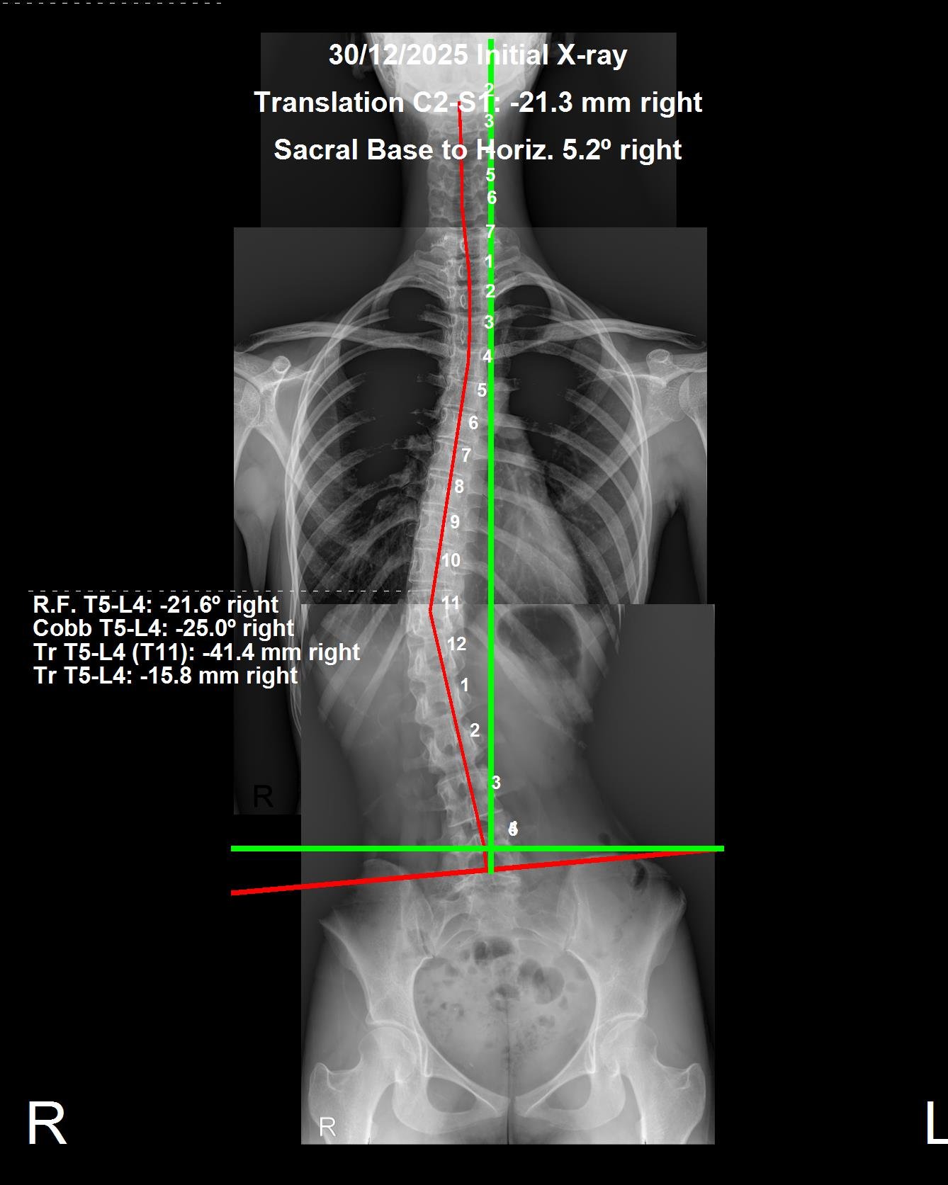
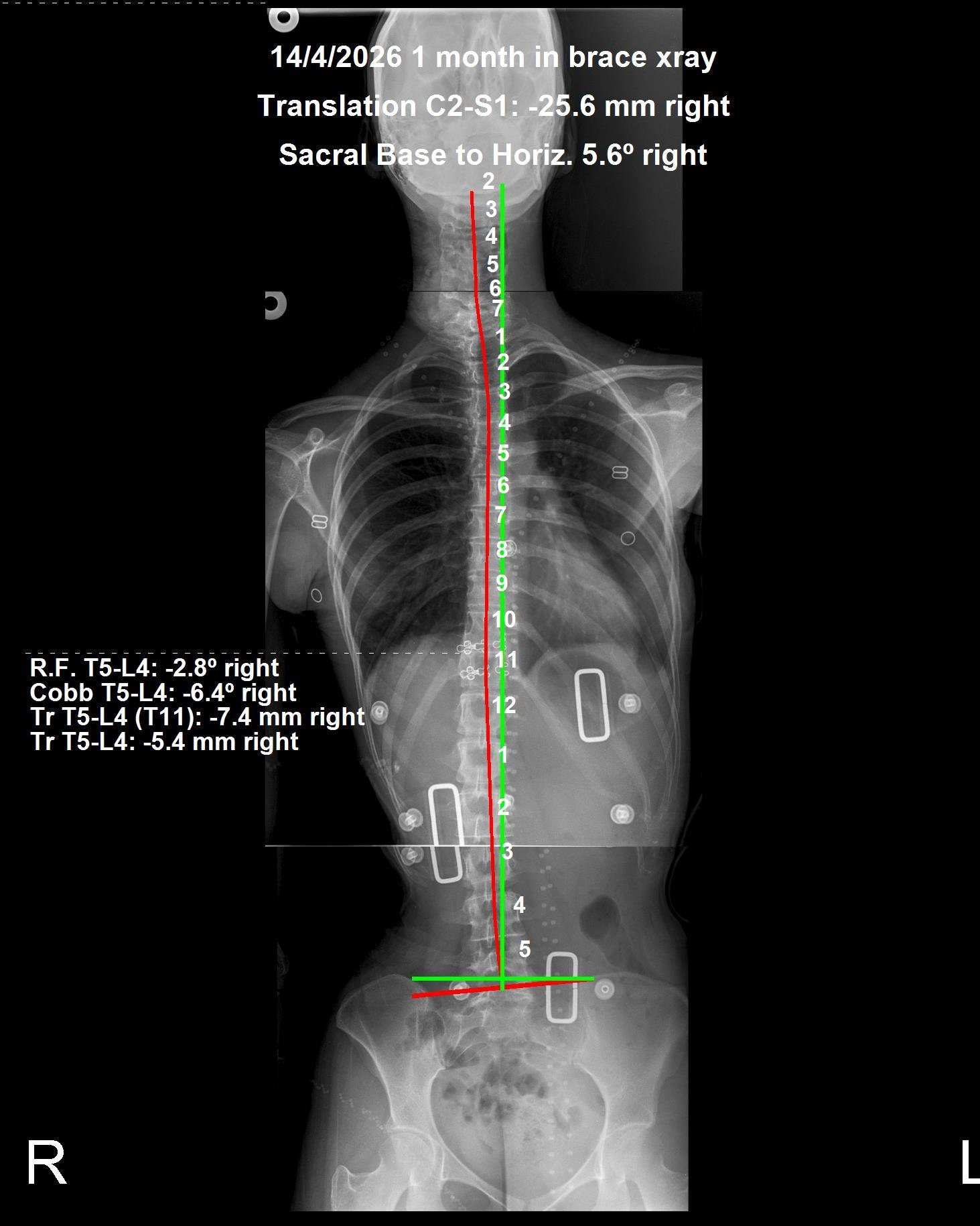
This patient presented to CBP Precision Spine Center in Muscat with a thoracolumbar scoliosis confirmed on PostureRay X-ray analysis. Pre-brace imaging documented a 25° Cobb angle with an apex translation of -41.4mm right at the T5–L4 spinal segment — a clinically significant curve requiring structural intervention. Following custom ScoliBrace fitting by Dr. Richard Marchetti, in-brace PostureRay X-ray at 1 month of wear demonstrated a Cobb angle reduction to 6.4° and apex translation correction to -7.4mm right — a 74% reduction in curve magnitude and an 82% reduction in translation within the first month of bracing alone.
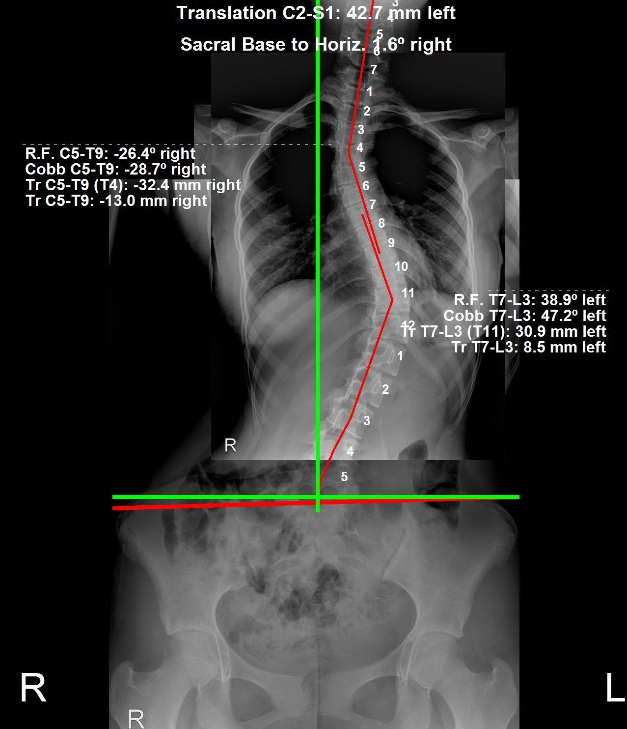
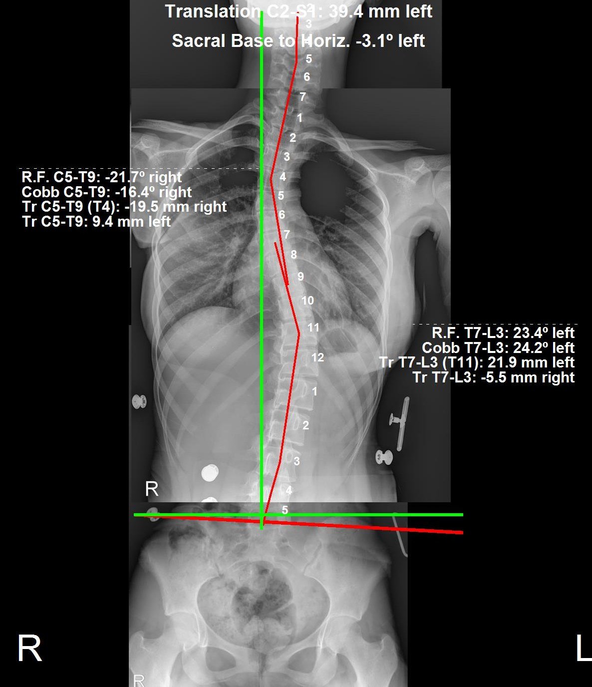
First diagnosed at age 10 and now 13, this patient reported no pain but was concerned about her scoliosis progressing. A previously prescribed brace was too flexible to control the curve. After 3 months in a custom ScoliBrace®, her main thoracolumbar curve reduced from 47.2° to 24.2° in-brace (a 49% correction) and her thoracic curve from 28.7° to 16.4°. She is very happy with her progress and is now beginning active CBP® treatment to further improve the curve.
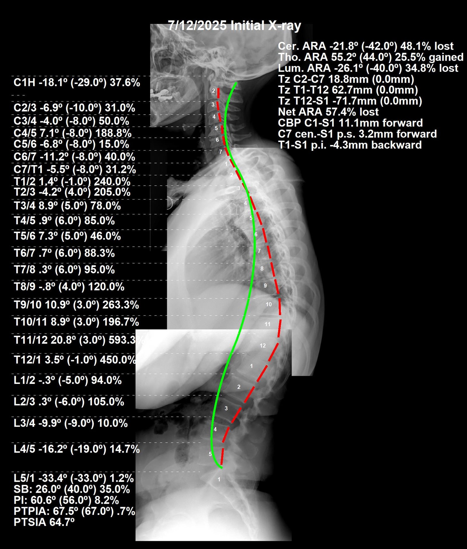
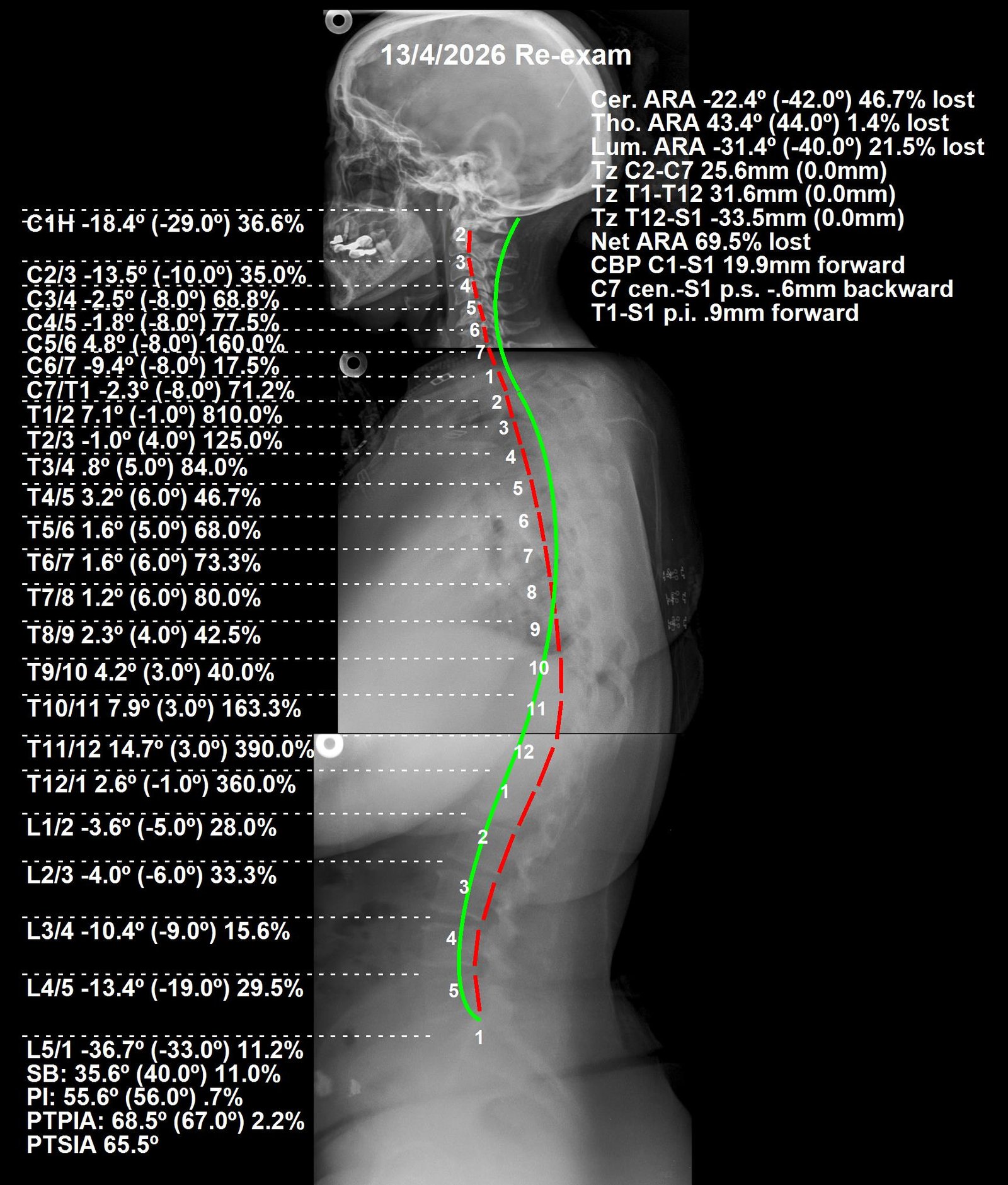
This patient presented to CBP Precision Spine Center in Muscat with chronic lower back pain and tingling radiating down both legs. PostureRay full spine X-ray analysis confirmed significant structural deviations across all three spinal regions — cervical, thoracic, and lumbar — each measurably outside the normal CBP® alignment values. The first 12 visits focused on pain management and nervous system stabilisation. The following 36 visits addressed structural correction using Chiropractic BioPhysics® mirror-image adjustments, region-specific extension traction, and a prescribed home rehabilitation programme. At 5 months, all three spinal regions showed measurable movement toward normal alignment on PostureRay follow-up X-ray, with full resolution of the patient's lower back pain and leg tingling.
| Region | Normal | Before | After |
|---|---|---|---|
| Cervical | 42° | -21.8° | -22.4° |
| Thoracic | 40° | 49.6° | 43.4° |
| Lumbar | 40° | -26.1° | -31.4° |
| Region | Normal | Before | After |
|---|---|---|---|
| Cervical | 0 mm | 18.8 mm | 25.6 mm |
| Thoracic | 0 mm | 62.7 mm post. | 31.6 mm post. |
| Lumbar | 0 mm | -71.7 mm post. | -33.5 mm post. |
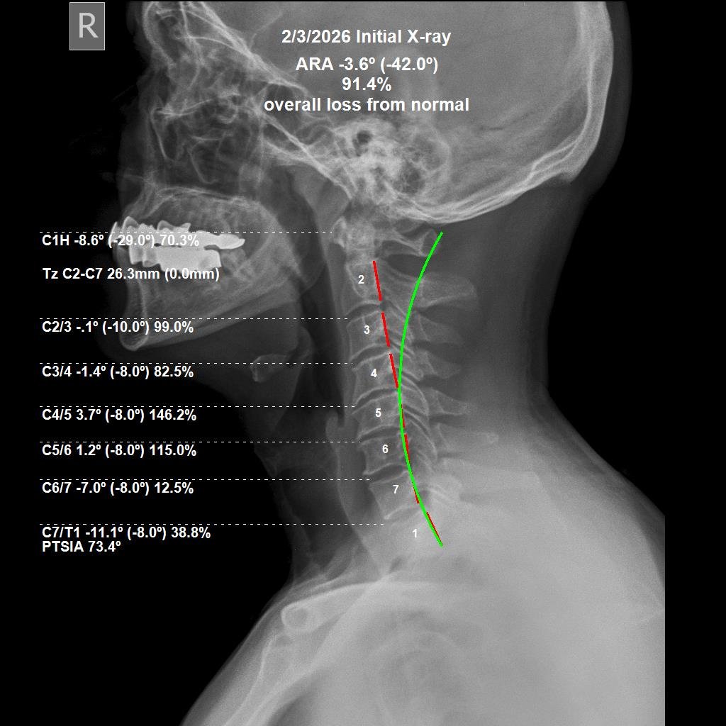
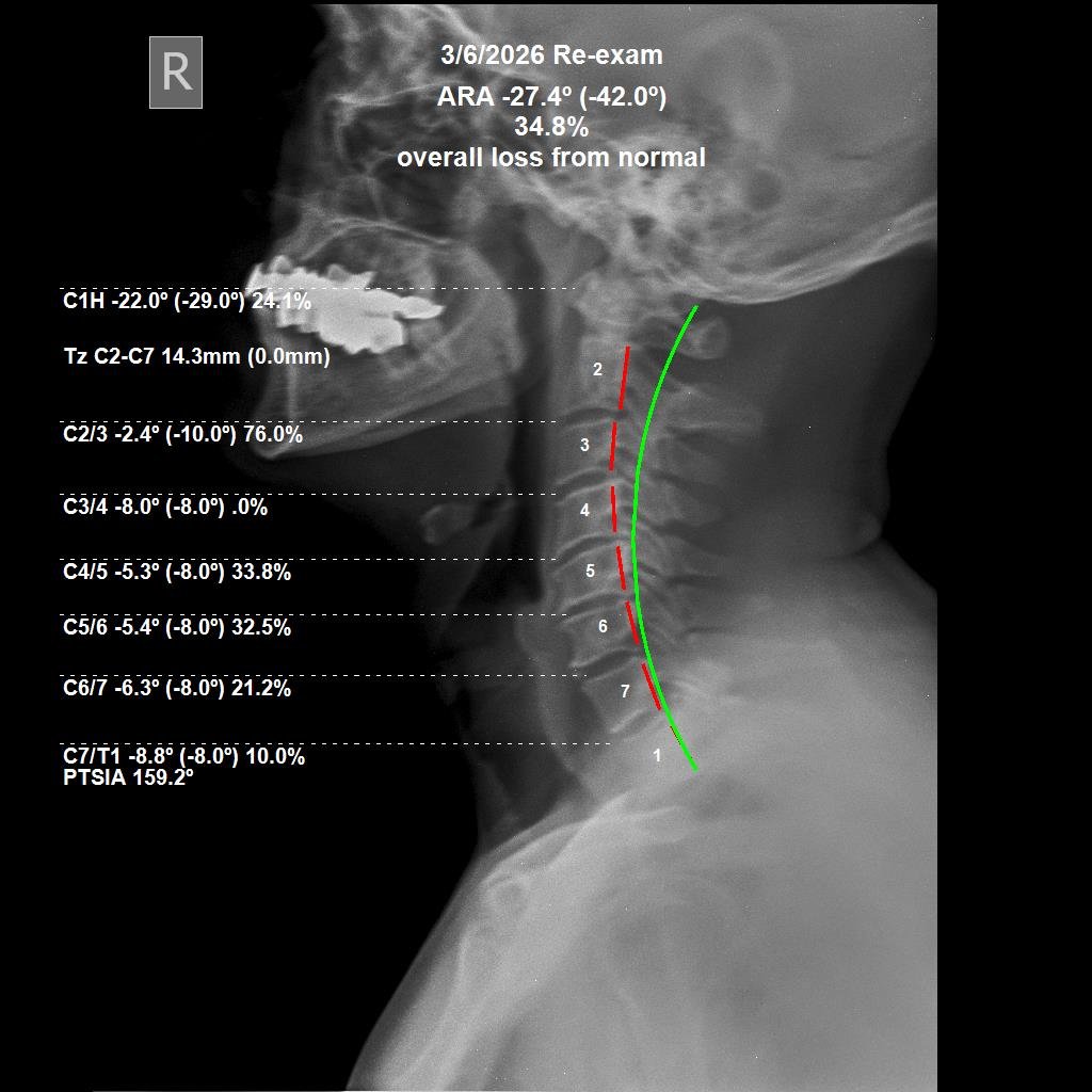
This patient came to CBP Precision Spine Center in Muscat not with pain — but with the self-awareness to get a postural and spinal check-up. To the eye, her posture looked close to perfect, with only minor visible deviations. But PostureRay X-ray analysis told a very different story. The cervical spine measurements revealed a 91.4% overall loss from normal alignment — a significant hidden structural deficit that carries long-term neurological and functional consequences regardless of how the patient looks or feels. Following 36 visits of Chiropractic BioPhysics® cervical structural correction — including mirror-image adjustments, cervical extension traction, and a home rehabilitation programme — the overall loss from normal was reduced to 34.8%. That represents a 56.6 percentage point improvement in cervical structural alignment in just 3 months. The patient reported feeling noticeably lighter, with reduced brain fog, improved flexibility, and stronger overall function — outcomes consistent with improved nervous system efficiency as the cervical spine moves toward its normal alignment.
| Measurement | Normal | Before | After |
|---|---|---|---|
| ARA C2–C7 | -42.0° | -3.6° (91.4% loss) | -27.4° (34.8% loss) |
| TZ C2–C7 | 0.0 mm | 26.3 mm | 14.3 mm |
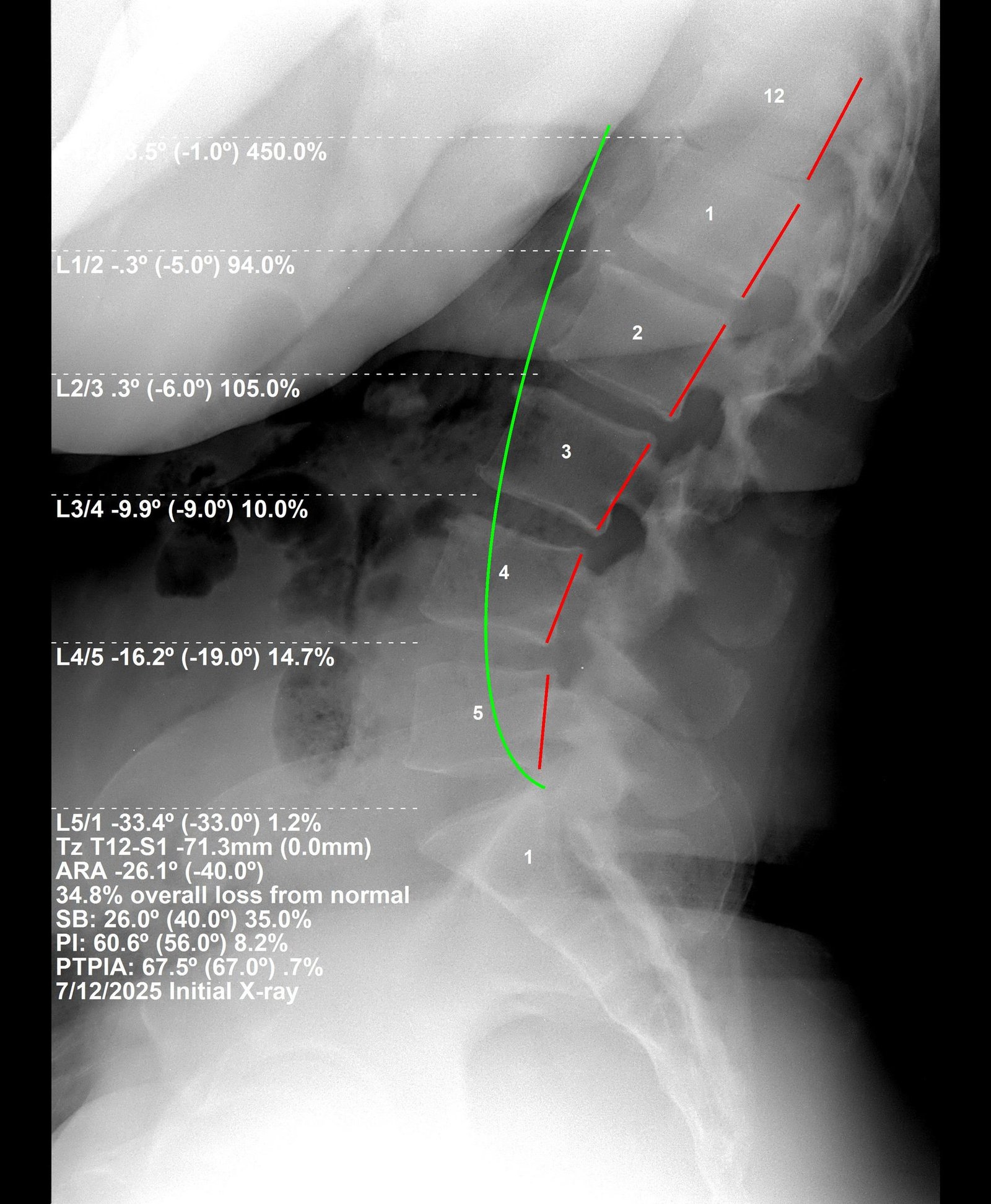
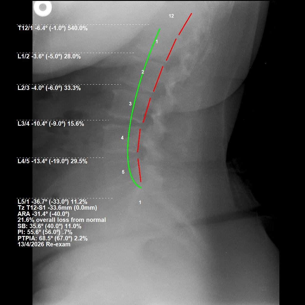
This patient presented to CBP Precision Spine Center in Muscat with tingling and numbness radiating down both legs — a common but serious symptom of lumbar structural deficit and its effect on the surrounding nervous system. PostureRay X-ray analysis confirmed the lumbar spine was operating at a 34.8% overall loss from its normal alignment, with significant posterior translational shift placing mechanical stress on the spinal cord and nerve roots. Following 36 visits of Chiropractic BioPhysics® lumbar structural correction — including mirror-image lumbar extension traction, region-specific drop adjustments, and a prescribed home rehabilitation programme — PostureRay follow-up X-ray at 4 months documented measurable structural improvement across both measurements. The overall loss from normal reduced from 34.8% to 21.6%, and the translational shift reduced by more than half. The patient reported complete resolution of tingling and numbness, the ability to pray without pain, and the capacity to walk significantly longer distances than before care.
| Measurement | Normal | Before | After |
|---|---|---|---|
| ARA L1–L5 | -40.0° | -26.1° (34.8% loss) | -31.4° (21.6% loss) |
| TZ T12–S1 | 0.0 mm | -71.3 mm post. | -33.6 mm post. |
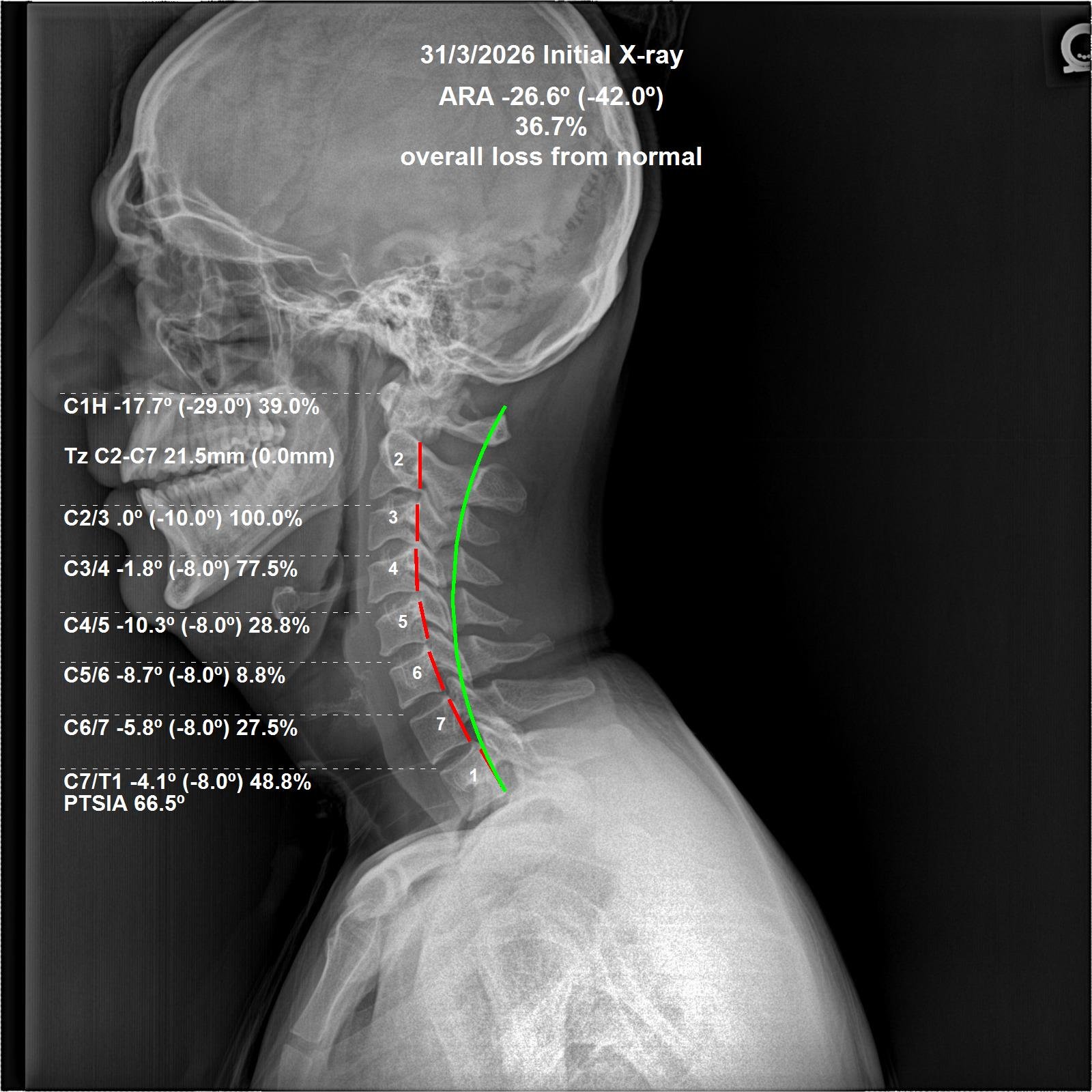
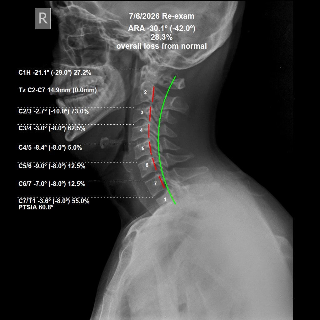
This patient presented to CBP Precision Spine Center in Muscat with neck pain, upper back pain, and additional lower back pain. PostureRay cervical X-ray analysis confirmed a 36.7% overall loss from the normal cervical curve — a structural deficit that places abnormal tension on the spinal cord and surrounding musculature, a common driver of both localised neck pain and referred pain patterns throughout the back. Following 36 visits of Chiropractic BioPhysics® cervical structural correction — including mirror-image adjustments, cervical extension traction, and a prescribed home programme — PostureRay follow-up X-ray at 3 months showed measurable improvement across both cervical alignment measurements. The overall loss from normal reduced from 36.7% to 28.3%, and the translational shift reduced from 21.5mm to 14.9mm. Notably, the patient's lower back pain resolved alongside the neck and upper back pain — a finding consistent with the global tension-releasing effect of restoring the cervical curve, even without direct lumbar intervention.
| Measurement | Normal | Before | After |
|---|---|---|---|
| ARA C2–C7 | -42.0° | -26.6° (36.7% loss) | -30.1° (28.3% loss) |
| TZ C2–C7 | 0.0 mm | 21.5 mm | 14.9 mm |
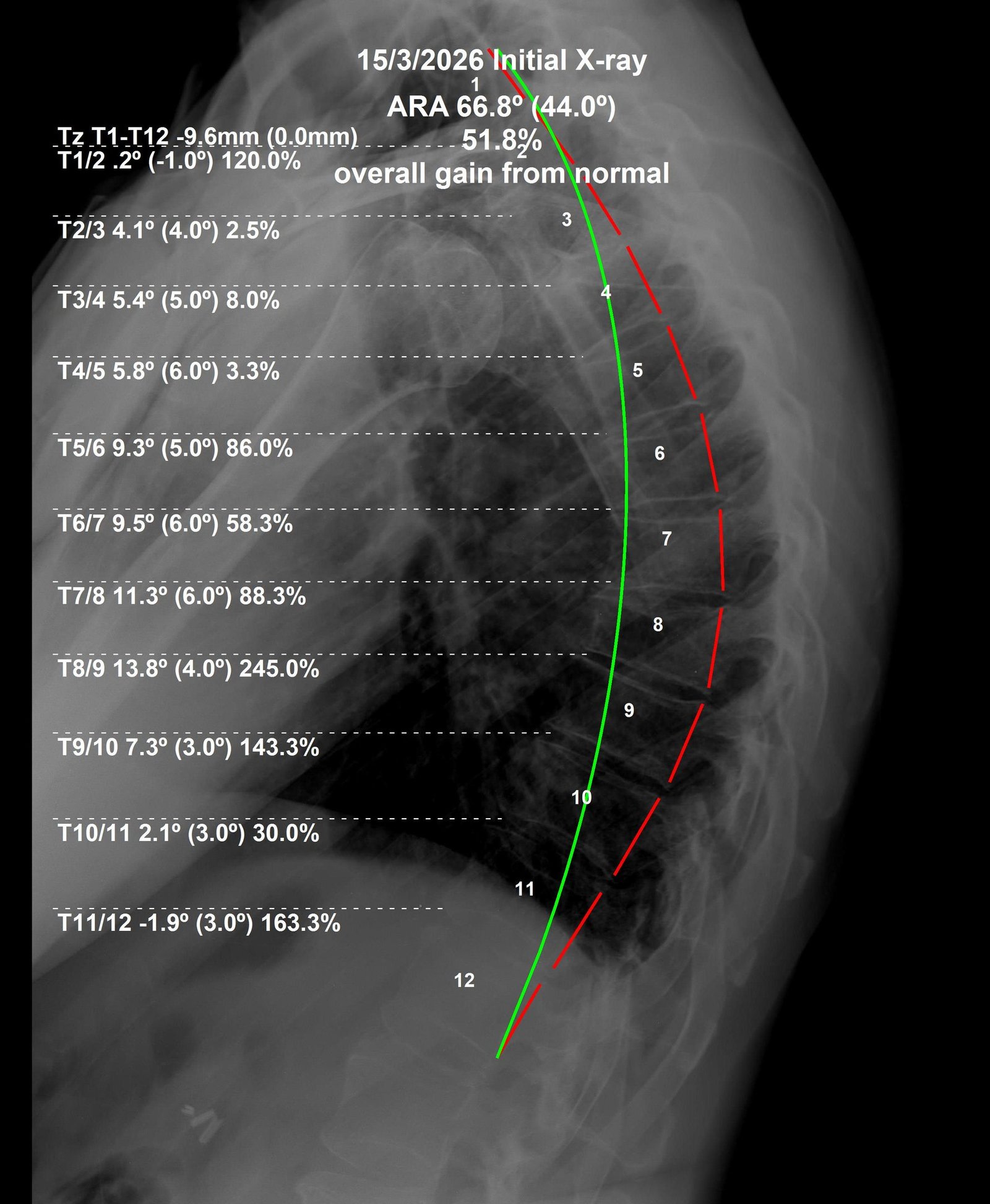
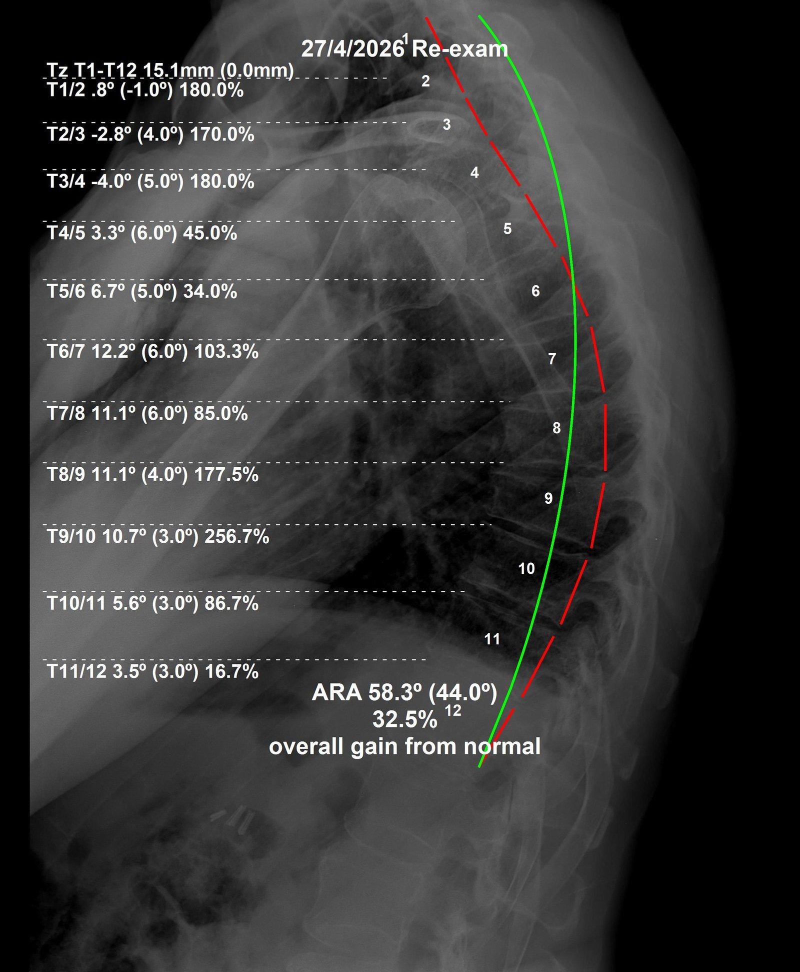
This patient travelled from France specifically to receive CBP® structural correction at CBP Precision Spine Center in Muscat, combining treatment with a holiday in Oman. He came in with no pain — his goal was postural improvement. PostureRay X-ray analysis revealed a diagnosis of Scheuermann's Kyphosis, with the thoracic curve measuring 66.8° — a 51.8% overall gain from the normal 44° thoracic arc, with an accompanying posterior translational shift. Despite the structural rigidity characteristic of Scheuermann's Kyphosis, PostureRay follow-up X-ray after just 2 months and 24 visits of Chiropractic BioPhysics® thoracic structural correction showed meaningful correction across both measurements. The thoracic ARC reduced from 66.8° to 58.3°, and the overall gain from normal reduced from 51.8% to 32.5% — a 19.3 percentage point improvement in 8 weeks. The patient had to return to France but plans to return to Muscat to complete a full 36-visit care plan and further the structural correction.
| Measurement | Normal | Before | After |
|---|---|---|---|
| ARA T1–T12 | 44.0° | 66.8° (51.8% gain) | 58.3° (32.5% gain) |
| TZ T1–T12 | 0.0 mm | -9.6 mm | 15.1 mm |
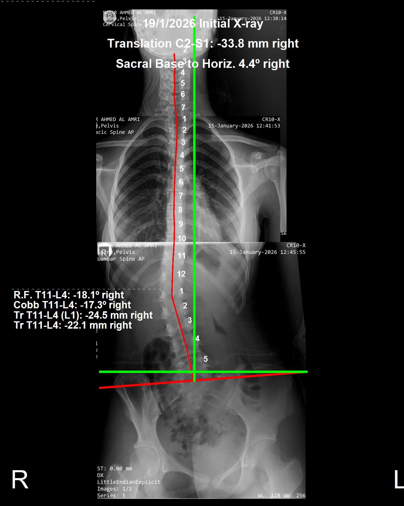
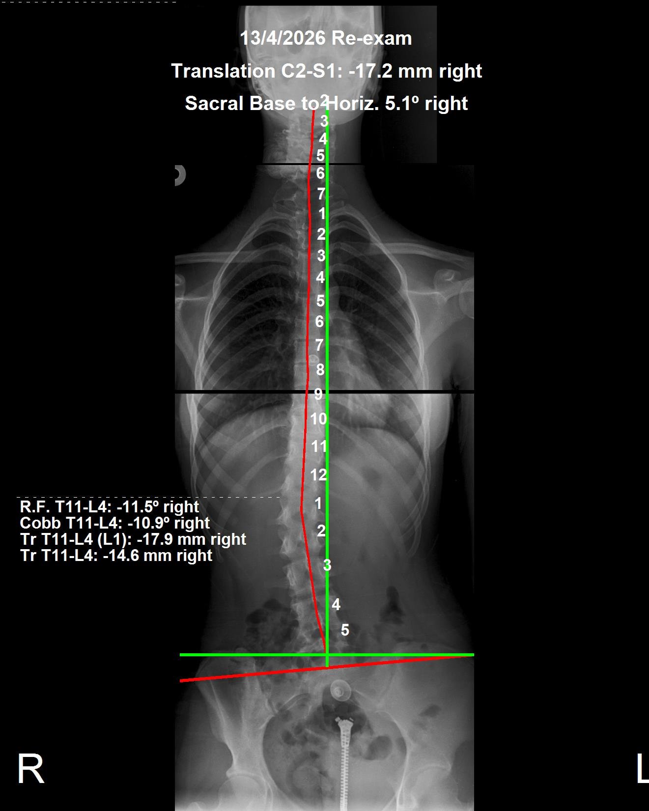
This patient presented to CBP Precision Spine Center in Muscat with a right thoracolumbar scoliosis confirmed on PostureRay X-ray analysis, along with leg shaking symptoms and numbness in the right foot — neurological signs consistent with mechanical nerve irritation from the spinal curvature and translation. Importantly, no ScoliBrace was used in this case. Correction was achieved entirely through the CBP® mirror-image traction and adjustment protocol: a series of region-specific extension traction setups designed to reverse the exact direction of the structural deviation, combined with a home rehabilitation programme. At 3 months and 24 visits, the Cobb angle reduced from 17.3° to 10.9° right — a 36.9% reduction — and the spinal translation reduced from -33.8mm to -17.2mm right, almost halving the lateral shift. The leg shaking stopped completely and the right foot numbness resolved. The patient elected to continue care given the degree of change achieved in such a short time.
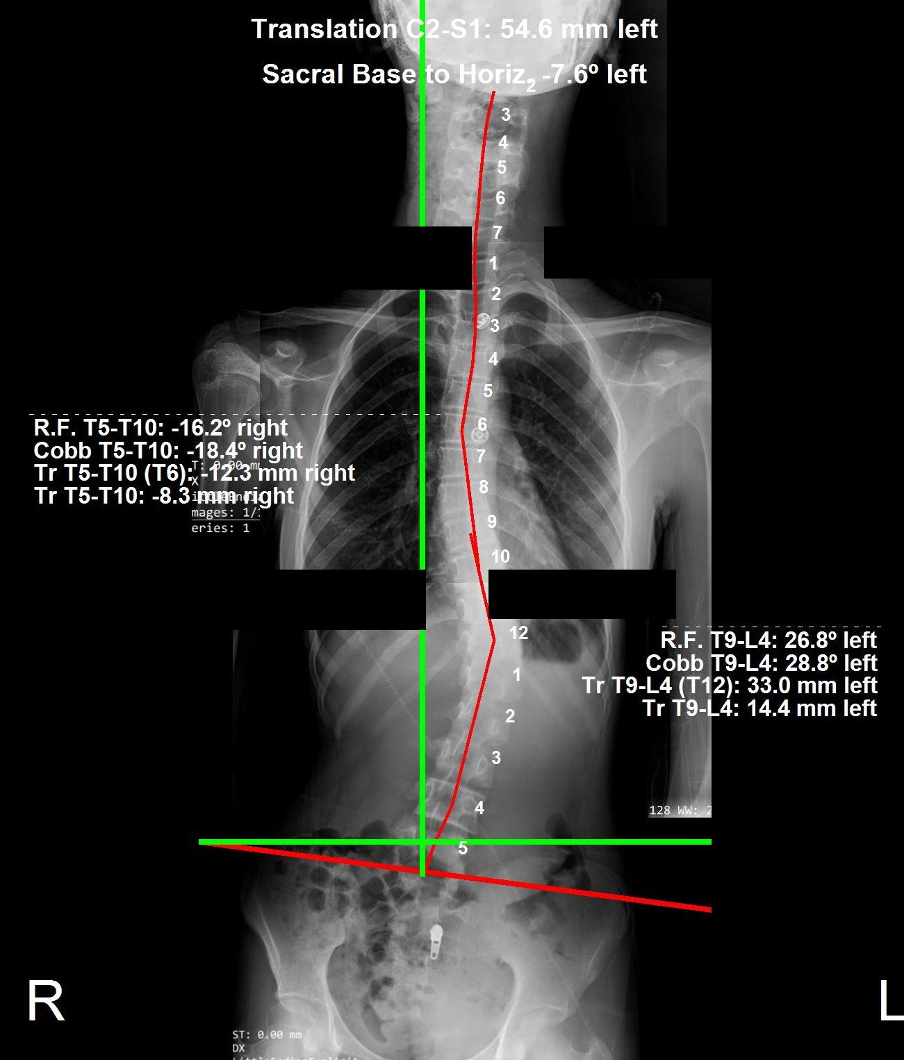
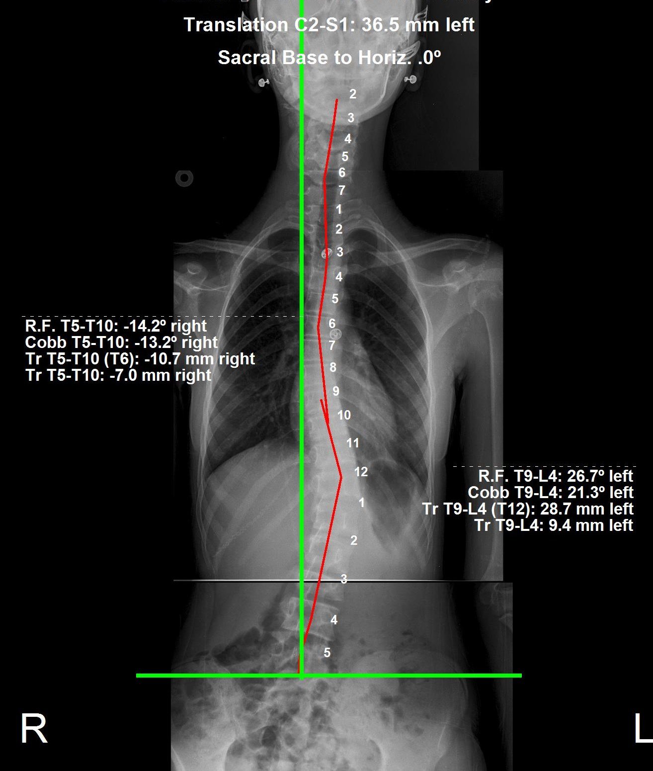
This patient noticed her posture had changed at age 13 — uneven shoulders that showed in the way she walked — and suspected scoliosis. Online exercises seemed to improve how she looked from the outside, but her spine stayed visibly curved. Timeline: the initial X-ray was taken at her first assessment in January 2026; her custom ScoliBrace® was not fitted until March 2026, and this follow-up was taken out of brace in June 2026, after about three months of bracing. It shows her main thoracolumbar curve reduced from 28.8° to 21.3°, her thoracic curve from 18.4° to 13.2°, and overall spinal balance (C2–S1 translation) improving from 54.6 mm to 36.5 mm.
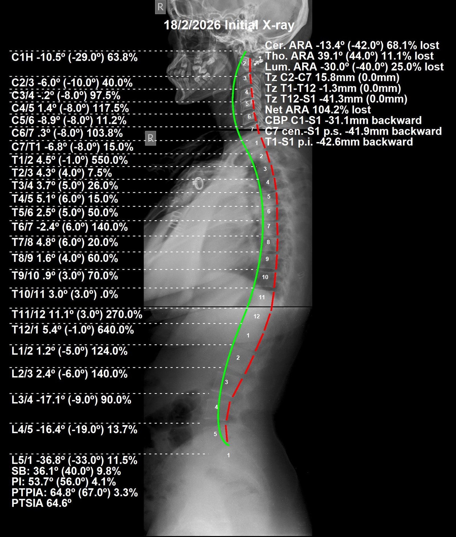
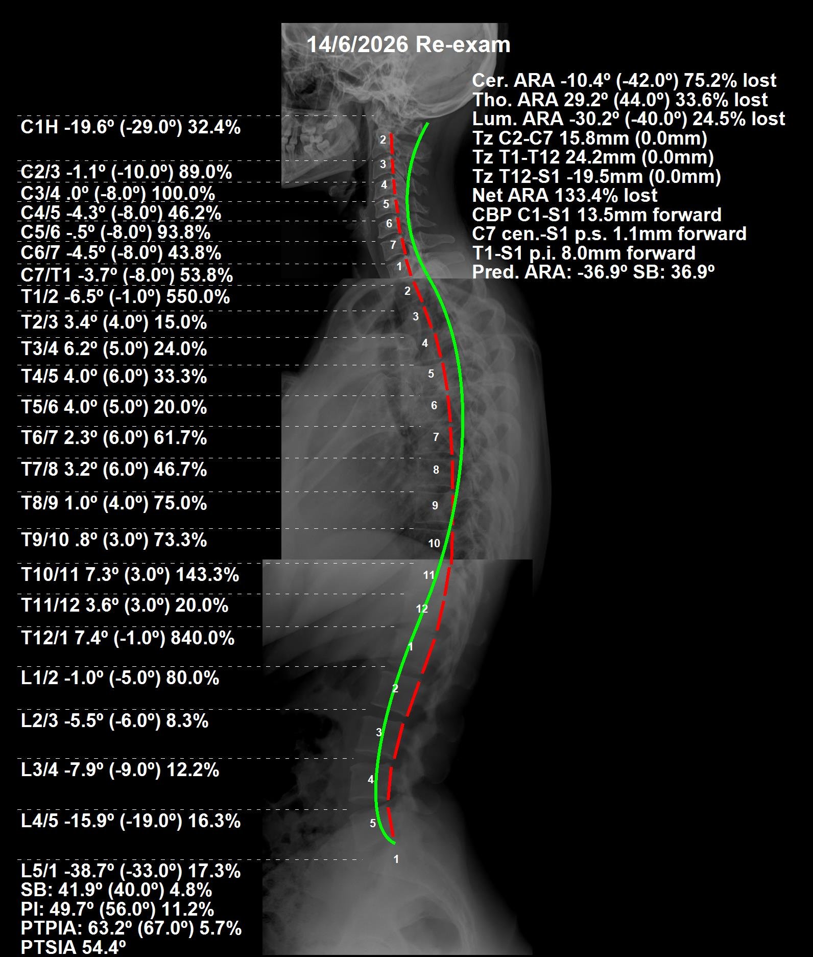
This patient presented to CBP Precision Spine Center in Muscat with radicular neck pain and lower back pain. PostureRay full spine X-ray analysis showed significant global sagittal imbalance, with the trunk carried well behind neutral — a T1-S1 sagittal offset of -42.6mm. Treatment used a Chiropractic BioPhysics® full spine protocol — mirror-image adjustments, region-specific extension traction, and a prescribed home rehabilitation programme. Across 36 visits over roughly four months, global sagittal balance was brought to near-neutral (T1-S1 +8.0mm — an ~81% reduction in offset), with lumbar translational displacement (T12-S1) reduced by roughly half. The patient's radicular neck and lower back pain resolved, leaving only minor residual hand symptoms after long workdays.
| Region | Normal | Before | After |
|---|---|---|---|
| Cervical | 42° | -13.4° | -10.4° |
| Thoracic | 44° | 39.1° | 29.2° |
| Lumbar | 40° | -30.0° | -30.2° |
| Region | Normal | Before | After |
|---|---|---|---|
| Cervical | 0 mm | 15.8 mm | 15.8 mm |
| Thoracic | 0 mm | -1.3 mm | 24.2 mm |
| Lumbar | 0 mm | -41.3 mm | -19.5 mm |